First Stage: Before

Develop early detection and diagnosis to understand the the underlying cause of stroke and other neurological conditions through novel minimally-invasive techniques and machine learning.

Research Projects

Magnetic Resonance Imaging for Idiopathic Intracranial Hypertension and Additional Cerebral Spinal Fluid Pathologies

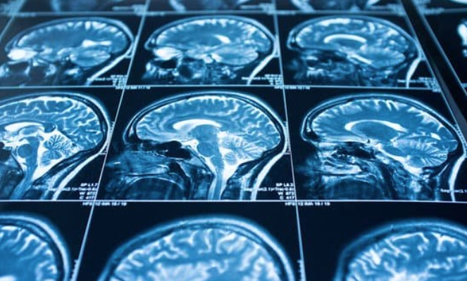
This study aims to gain an understanding of the cerebrovascular factors that contribute to idiopathic intracranial hypertension (IIH) and additional cerebrospinal fluid (CSF) pathologies using specialized magnetic resonance imaging (MRI) techniques to ultimately improve patient outcomes. Eligible patients are diagnosed with IIH and have yet to undergo interventional treatments. 
Model Seeding: Cerebral aneurysm risk stratification using hemodynamic modeling and endothelial genetics

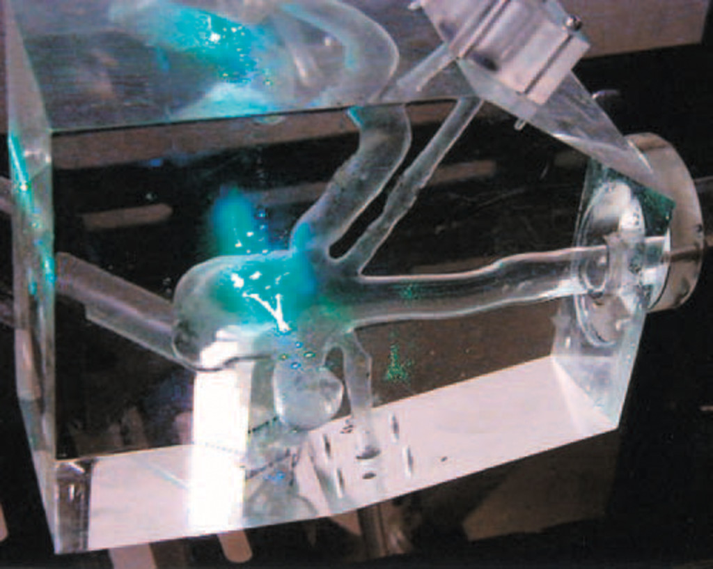
The Cerebral aneurysm risk stratification using hemodynamic modeling and endothelial genetics (Model Seeding) project utilizes patient-specific3D-printed models of cerebral aneurysms seeded with vascular endothelial cells to uncover the relationship between hemodynamic and genetic variables in the context of intracranial aneurysm (IA) formation, growth, and ruptureComputational Fluid Dynamics (CFD) simulations are created for a variety of aneurysms of different sizes and configurations to obtain accurate maps of hemodynamic stresses. Then, the aforementioned 3D-printed, patient specific, “endothelialized” models is exposed to flow in order to recreate the hemodynamic conditions simulated in the CFD models. After exposure to flow, the model’s endothelial cells are biopsied according to areas of hemodynamic interest highlighted by the previously made CFD simulations. mRNA and protein quantities of key biological factors produced by these endothelial cells in response to different patterns of hemodynamic stresses are measured and related to the magnitude of the mechanical stimuli calculated by CFD simulations in those same anatomic locations. This project is supported by an Institute of Translational Health Sciences Research Innovation Award.  

Endovascular biopsy for the investigation of the genetics and basic biology of diseases of the brain and its blood vessels

With the support from the BEE Foundation, our Endovascular Biopsy project investigates the genetics and molecular biology of patients with diseases of the brain and its blood vessels. For the project, we have optimized a novel, minimally invasive method of collecting subjects’ cells from areas of pathology (such as aneurysms and arteriovenous malformations [AVM’s]) during endovascular treatment. Medical devices that are used in surgery come into contact with blood vessel cells. Cells that have lodged onto the medical devices are isolated via Fluorescence Activated Cell Sorting (FACS) and stored. After isolation, we perform targeted transcriptome measurements via next generation, single-cell RNA sequencing. Ultimately, we will be able to understand the genetic underpinnings of cerebrovascular diseases. This enhanced understanding could lead to novel therapies and better treatment outcomes  

GeneFlow: Genetic and hemodynamic parameters of intracranial aneurysm treatment outcomes, formation, and rupture

This study, funded by the University of Washington Office of Research Royalty Research Fund (RRF), is an expansion of our Model Seeding and Endovascular Biopsy projects. We seek to improve the accuracy and clinical translation of our models by inputting intraoperatively acquired, patient-specific flow parameters and vascular endothelial cells. During endovascular treatment of an aneurysm Doppler flow wires will be used to measure the physical parameters of blood flow around the aneurysm and medical devices will be collected to isolate and grow vascular endothelial cells from the pathologic vascular tissue. These flow parameters will be used in computational fluid dynamics (CFD) simulations and the collected endothelial cells will be used to line the models of our 3D-printed, patient-specific aneurysm models.

Computational fluid dynamics of Alzheimer’s disease

The cerebrospinal fluid (CSF) in patients exhibiting Alzheimer’s disease (AD) is observed to contain plaques and tangles of AD-related molecules that alter brain tissue. Prior studies found that that a CSF shunt placement reduced the presence of AD-related proteins. Whypothesize that computational method of determining the CSF-related stresses on surrounding brain tissue would permit noninvasive, early diagnosis of AD and mild cognitive impairment (MCI) 

Come Meet Us Up Close

Idiopathic Intracranial Hypertension

Magnetic Resonance Imaging for Idiopathic Intracranial Hypertension and Additional Cerebral Spinal Fluid pathologies


Aneurysm

Model Seeding: Cerebral Aneurysm risk stratification using hemodynamic modeling and endothelial genetics

Aneurysm and Arteriovenous Malformation

Endovascular biopsy for the investigation of the genetics and basic biology of diseases of the brain and its blood vessels

Aneurysm and Arteriovenous Malformation

Endovascular biopsy for the investigation of the genetics and basic biology of diseases of the brain and its blood vessels

Alzheimer

Computational fluid dynamics of Alzheimer's disease